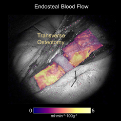
An overlay of the endosteal blood flow map on a grayscale white-light image of the surgical field.
Senior Scientist and Assistant Professor of Surgery Jonathan T. Elliott, along with Orthopedic Surgeon Leah Gitajn and Engineering Professors Shudong Jiang and Brian Pogue published a Letter in Journal of Biophotonics this month describing a new method of imaging blood flow in the bone that may be able to tell surgeons whether bones involved in complex fractures—such as high-force trauma caused by motor vehicle accidents—are adequately supplied by blood vessels in the surrounding tissue (periosteum) or from within the bone (endosteal route), both of which are present in health bone.
Parametric images of periosteal blood flow, endosteal blood flow, total bone blood flow and fraction of endosteal-to-periosteal flow were obtained by applying the hybrid plug/compartment (HyPC) kinetic model on a pixel-by-pixel basis.
Enabling discrimination between healthy and damaged bone intraoperatively could help surgeons during debridement (removal of dead bone), lowering the incidence of post‐operative complications from infection and poor healing. Furthermore, the ability to quantify periosteal and endosteal blood flow could help surgeons avoid further compromising bone blood supply by helping them choose whether to reinforce the remaining bone using a long nail placed inside the bone or using a plate-and-screw method.
Read the full Letter here (requires access to subscription).