The Orthopaedic Translational Research Laboratory (OTEL) is federally-funded to conduct research in several areas of inquiry. Some of our ongoing projects are summarized here, and reflect the work of team members and collaborators.
Click on each Project Title to see a Summary:
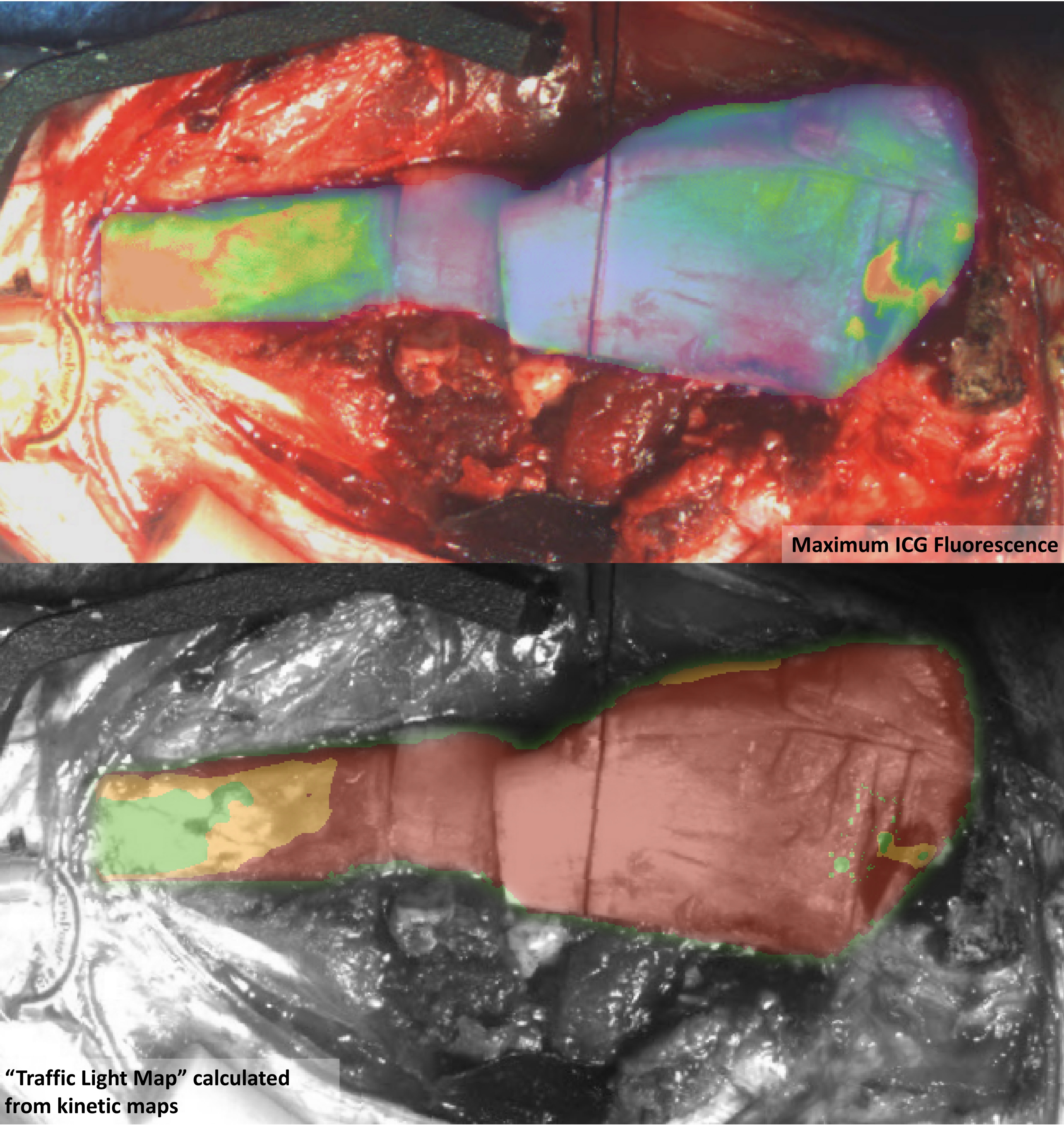
Fluorescence map of tibia fracture with significant periosteal stripping (top) and traffic-light map indicating predicted bone viability (bottom).
Significance: Infection following bony fracture is one of the most prevalent and challenging complications in trauma patients with an estimated annual cost of $35 billion in the US. Because traumatic fractures are often contaminated with foreign bodies, and they frequently require metallic implants for definitive fixation, they are inherently prone to infection. About 60% of high-energy fractures will develop infection, and about 30% of infections are not treated successfully. The mainline treatment for bony infection is thorough debridement of devitalized and infected bone.
Project Aims: This project involves developing the concept of fluorescence-guided debridement (FGD)—the use of imaging-based fluorescence contrast to guide debridement of open fractures contaminated with devitalized and infected tissue.
Approach: In the heart of our Preclinical/Translational Biosafety Level 2 laboratory, using clinically relevant rodent and large-animal models of high-energy and low-energy open fracture and bone infection, we are optimizing the selection and timing of fluorescent probes and demonstrate the efficacy of FGD for managing deep wound infection.
Impact: Open fractures are associated with a 30% infection rate which can have devastating downstream consequences including unplanned amputation, and successful completion of this project could revolutionize clinical management of orthopaedic trauma.
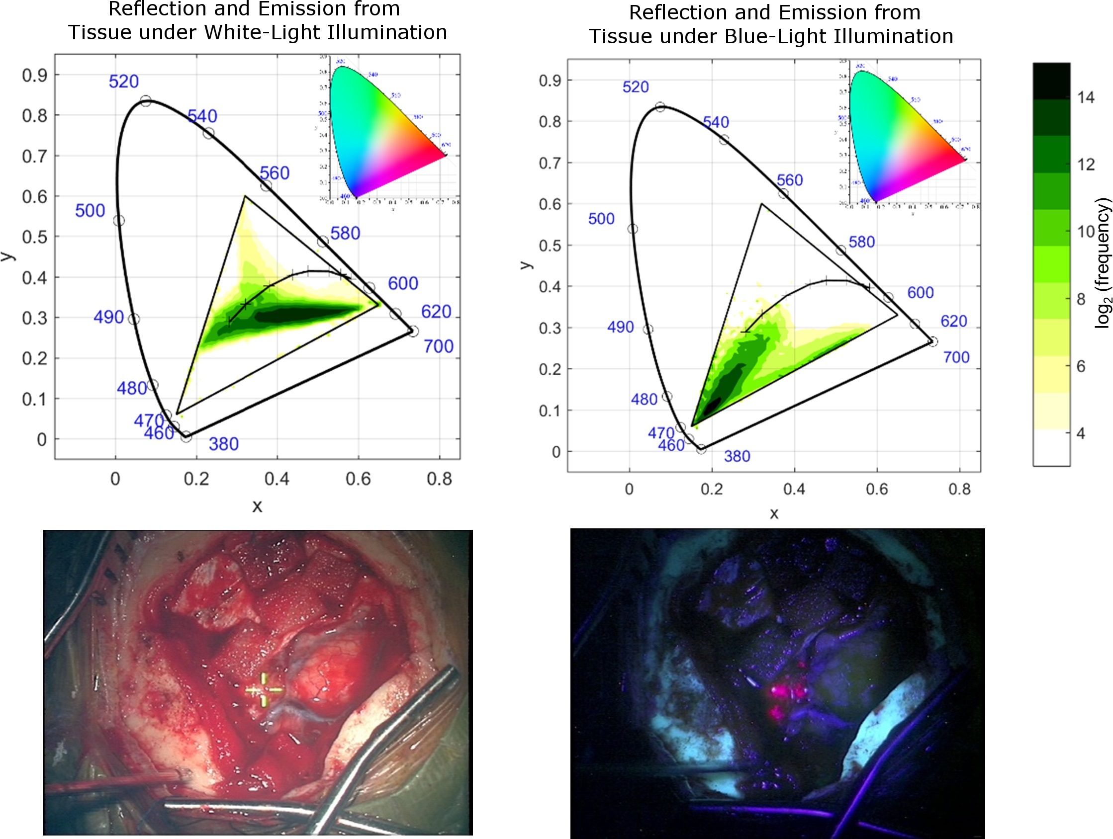
Density maps (top) showing the occurance of pixels of a particular chromaticity in white-light mode (bottom, left) and blue-light mode (bottom, right) during brain resection.
Significance: Surgical microscopy still involves the direct observation of light through an objective lens, relay system and eyepiece magnifier—essentially unchanged since the early 20th century. A new type of system is needed to fully leverage the current advances in fluorescence guided surgery (FGS), which is gaining wide acceptance in the surgical oncology community. In particular, the ability of FGS to enhance high-grade gliomas through 5-aminolevulinc acid (5-ALA) induced protoporphyrin IX (PpIX) is improving extent of tumor resection.
However, gains are not being realized in other more treatable low-grade glioma (LGG) tumors because the levels of PpIX are much lower. The development of more sensitive camera technology for PpIX, applying spectral sitting, and using targeted "smart" probes, many of which emit infrared light, means a different way of visualizing fluorescence. In these cases, fluorescence must be captured and redisplayed on a monitor or in a heads-up display (HUD)/augmented reality (AR) platform. This requires the fusion of fluorescence data—represented by a parametric map of colors that correspond to particular intensity values—with the visual representation of the surgical field.
Project Aims: This project involves (1) Building and evaluating strategies using a pulsed/CW dual-mode light source that can allow either direct 5-ALA visualization or white-light visualization with secondary monitor 5-ALA/PpIX map, to improve the visibility of structures and anatomy in the normal brain without reducing perceived brightness of the tumor, (2) Optimizing the color map and transparency function used when fusing white-light and fluorescence images together, so that fluorescence information is easily and accurately communicated, and features in the white-light image are not obscured.
Approach: Hardware characterization in preclinical models and usability studies in the clinical operational environment are complimented by an exploration of themes in visual perception, philosophy and psychology.
Impact: The optimal way of combining this information has not yet been elucidated, despite the fact that data visualization is the essential step translating improved imaging technology into surgeon decision-making and actual patient outcome. We are developing a platform that will allow a surgeon to have an identical experience to using a microscope, but instead interface with a stereo multichannel imaging system.
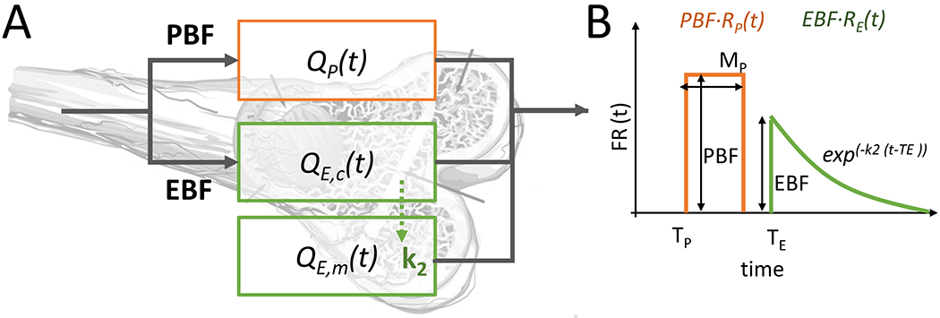
(A) A diagram showing the compartments of the HyPC model (B) The flow-scaled aggregate impulse residue function used in the HyPC model, showing the parameters recovered (source: J Biomed Opt).
Significance: To directly measure the perfusion of tissue—how much volume of blood is delivered to a unit mass of tissue per minute—an imaging technique called "tracer kinetic analysis" can be used. Simply put, this approach introduces a contrast agent into the subject intravenously and then measures both the concentration of dye as a function of time circulating in the arterial system, and the time-varying concentration of dye in the tissue of interest. These two functions are related by a convolution: the arterial input function (AIF) convolved with a tissue-specific impulse function (IRF) is equal to the tissue dye concentration. The IRF can be modeled (kinetic modeling) or it can be deconvolved, to calculate physiological values like blood flow (perfusion), permeability (leakiness), blood volume, retention.
Project Aims: The purpose of this project is to support some of the other projects through the refinement of hardware and software. Aim 1 is to build an indocyanine green "pulse dye densitometer" that can non-invasively measure the circulating arterial concentration of dye. Aim 2 is to continue to develop parametric or model-based approaches to extracting blood flow parameters, with a focus on bone-specific models like the hybrid plug-flow compartment (HyPC) model [PMC7331892]. Aim 3 is to develop nonparametric methods that are rapid and correlate to proximal clinical outcomes even if they don't lend to a physical or physiological representation. Aim 4 is to engage in partnership with industry leaders in ICG-based intraoperative imaging to integrate AIF-acquisition hardware into clinical devices.
Impact: We've demonstrated the ability to quantify bone blood flow [PMC7331892] with first-pass kinetics and shown how important accounting for the arterial input function (AIF) is across all ICG dilution applications [PMC7282620].
The current opioid epidemic is a rapidly growing public health crisis in the US and Canada. In 2014, the almost 30,000 opioid-linked overdose deaths in the US was essentially equal to the number of fatal car accidents for the same year. I am leading a project to develop a unique imaging approach to predict, diagnose and help treat opioid use disorder. The goal of the project is to develop a targeted fluorescence imaging approach using labeled opioid agonists and antagonists to evaluate the changes in mu opioid expression receptor (MOR) that occur in the spiral ganglion of the ear. Ototoxicity and hypersensitivity are seen in ENT clinical practice associated with opioid abuse and risk of overdose. This leads us to hypothesize that changes in receptor behavior in these ganglia will correlate to progression and recovery in OUD. Through the development of this technology, we hope to provide a tool to understand the physiological progression of opioid addiction, both in basic science research, as well as in the administration of effective and objective opioid replacement therapy and addiction recovery.
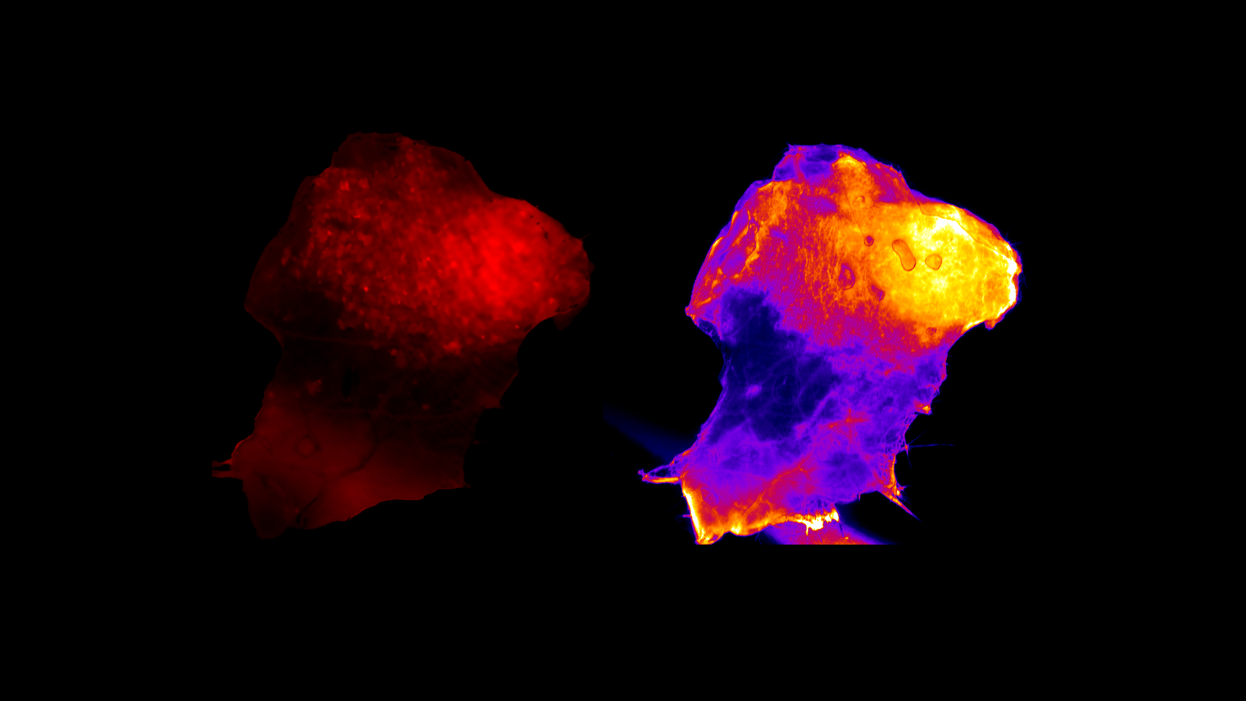
Bioluminescence detected from ovarian carcinoma cells on a tissue sample of mouse peritonium (left) and the ABY-029 fluorescence enhancement detected using wide-field imaging (right).
In collaboration with the Optics in Medicine Lab at Thayer School of Engineering at Dartmouth, we have developed and evaluated a targeted fluorescent probe—ABY-029—in a series of preclinical and clinical trials to detect EGFR-positive tumors in vivo. The initial clinical trials used microdose levels of agent, and therefore, our lab designed and built specialized imaging equipment that could retrofit onto clinical operating microscopes to enable sub-nanomolar sensitivity to the ABY-029 dye.
In addition to three clinical trials (glioma, head-and-neck, and sarcoma) the multi-institutional team is exploring the use of combinations of 5-ALA and ABY-029 in the detection of cancer, and preparing to advance the ABY-029 molecule into the next phase of clinical drug evaluation (Phase 1).
The overall goal of this project is to develop hardware and analysis software to measure the changes in signal and corresponding concentration changes of hemoglobin and deoxyhemoglobin during traumatic injury.
Two main projects use this technology but have very different applications:
The first project, led by PI Dr. Norman Paradis, and in Collaboration with Profs. Vikrant Vaze and Ryan Halter, is developing a system to provide early detection of hemorrhagic shock. In a mass casualty situation, such as a large bomb detonation or natural disaster, there are three types of patients: 1) the ones who are in really bad shape and it's fairly obvious, 2) the ones who have minimal injuries and will live regardless of what the interventions are, and 3) the ones who look like they aren't too bad, but then deteriorate. Our project intends to develop a system to identify the 3rd group of patients. Existing technologies have poor performance to identify group #3. Rigorous evaluation of their predictive value in this context has either been unsuccessful or unreported. Instead of taking a "single-sensor-single-location" approach, our method combines sensing technologies in a multiplex approach that could potentially outperform single sensor systems and bedside clinicians.
The second project, a collaboration between Prof. Elliott and Dr. Leah Gitajn, has built and tested a system capable of measuring tissue spectroscopy to detect tourniquet-associated injury during extremity orthopaedic surgery. In severe traumatic fracture, the use of tourniquet is thought to contribute to post-operative complications by depriving tissue of oxygen delivery. However, the mechanisms and exact relationship between oxy- deoxyhemoglobin, cytochrome c oxidase and clinical outcomes has not been well-studied. This project is improving our understanding of how tourniquet can be more judiciously used in certain patient populations without exacerbating injury.