Two New Publications on Bone Imaging
September 15, 2020
The Bone Imaging Team—a collaboration between the DS-GL, Department of Orthopaedics and Thayer—has published two important papers this summer that move the field of dynamic bone imaging forward, demonstrating cutting-edge techniques and future ideas that will enhance the quantative aspect of methodologies and provide possible clinical directions for future research.
The first paper, entitled "Intraoperative fluorescence perfusion assessment should be corrected by a measured subject-specific arterial input function" (https://doi.org/10.1117/1.JBO.25.6.066002) demonstrates the effect of variations in the arterial input function on the resulting qualitative and quantitative interpretations of fluorescence imaging during surgery. According to Prof. Jonathan Elliott, first author of the paper: "These effects propogate into observable parameters used in fluorescence imaging either implicitly by viewing the image or explicitly through intensity fitting algorithms, and should be corrected by patient-specific arterial input functions"
The second paper, entitled "Perspective on optical imaging for functional assessment in musculoskeletal extremity trauma surgery" (https://doi.org/10.1117/1.JBO.25.8.080601) is a Perspective paper highlighting the current challenges in assessing bone and tissue perfusion/viability and the potentially high impact applications for optical imaging in orthopaedic surgery. "For orthopaedic surgery, real-time data regarding bone and tissue perfusion should lead to more effective patient-specific management of common orthopaedic conditions, requiring deeper penetrance commonly seen with indocyanine green imaging," asserts Dr. Leah Gitajn, corresponding author of the paper.
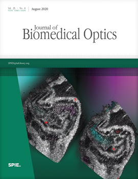
Both papers can be read in the August 2020 issue of Journal of Biomedical Optics.
← return to news
